
Photographic technique · visible fluorescence under UV excitation
UV-Induced Visible Fluorescence Photography (UVIVF), also called Ultraviolet Fluorescence Photography, records visible emission from UV-excited materials. Selective glow transforms flowers, minerals and ordinary surfaces while leaving other regions dark.
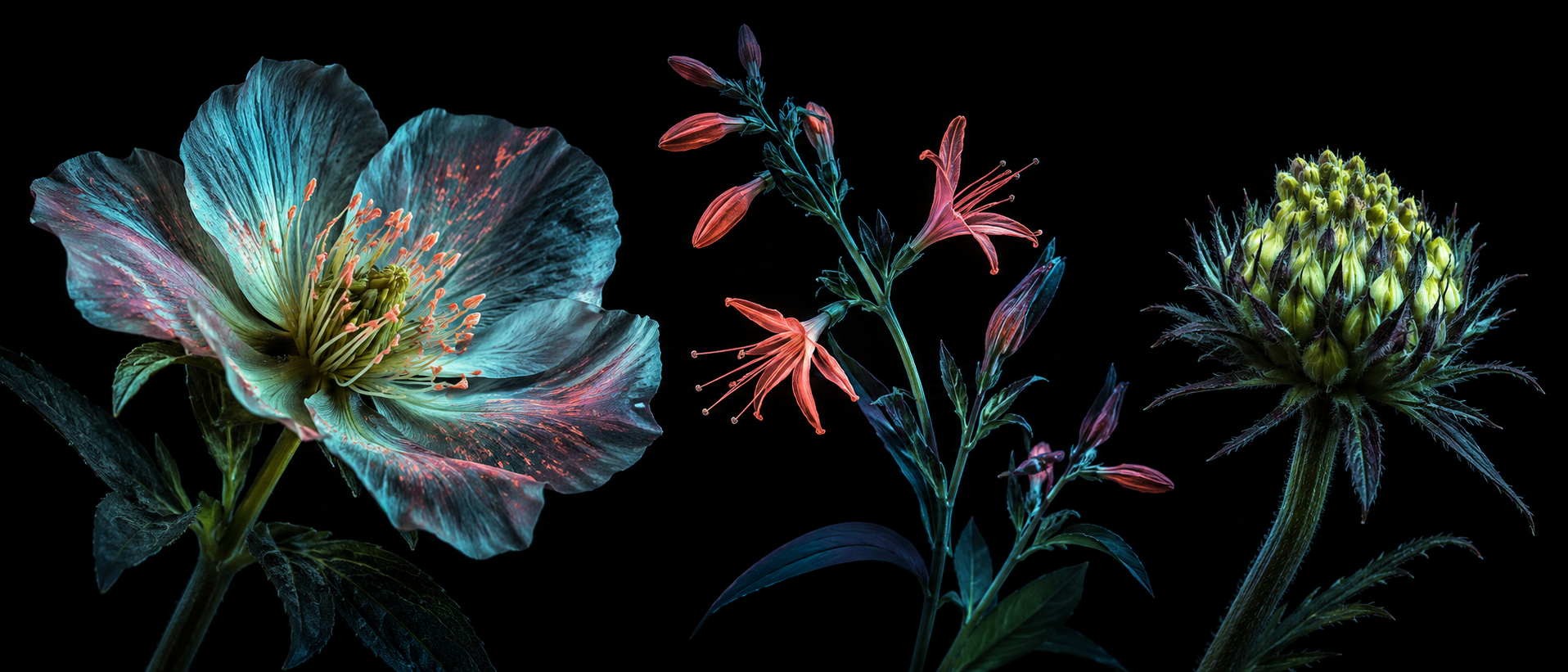
A visual introduction
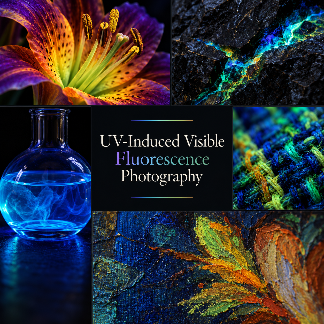
Seven original AI-generated AOI interpretations: one dedicated banner, one centrally titled collage and five standalone visual studies. Colors and material responses are illustrative, not measured fluorescence or diagnostic evidence.
Select an image for a closer look. Use the thumbnails, arrow keys or a swipe to compare examples. Zoom in to inspect details, or share the image that inspires you.
Creative toolbox · take this style further
Choose an input and a destination. Search sends your words to the chosen site; Copy & open puts them on your clipboard to paste there.
Create with AI
Turn your chosen input into an image prompt or a new creative direction.
ChatGPT
Copy your input and paste it into a ChatGPT conversation or image request.
Copy & open ChatGPT (new tab)Claude · Anthropic
Develop an image prompt or explore the style in a new Claude conversation.
Copy & open Claude · Anthropic (new tab)Midjourney
Paste your input into the Imagine bar on the Create page.
Copy & open Midjourney (new tab)Adobe Firefly
Copy your input, open Firefly, and paste it into the prompt field.
Copy & open Adobe Firefly (new tab)Leonardo.Ai
Copy your input, open Leonardo, and paste it into the generation field.
Copy & open Leonardo.Ai (new tab)Image search
Find visual references. Short, concrete keywords usually work best.
Lexica
Search AI images and prompts using your selected input.
Search with this input Lexica (new tab)Pexels
Search the photo and video library. Short keywords often work best.
Search with this input Pexels (new tab)Pixabay
Search photos, illustrations and other images.
Search with this input Pixabay (new tab)Unsplash
Search photographs using the style name or your own keywords.
Search with this input Unsplash (new tab)Wikimedia Commons
Search photographs, artworks and historical images. Check each file for its source and reuse terms.
Search with this input Wikimedia Commons (new tab)Art & research
Explore artists, collections and the ideas behind a style.
Wikipedia
Search for background reading. A style may not have its own article.
Search with this input Wikipedia (new tab)WikiArt
Copy your input into WikiArt’s search to explore artists and artworks.
Copy & open WikiArt (new tab)Europeana
Paste your keywords into the search to explore European museum, library and archive collections.
Copy & open Europeana (new tab)Inspiration
Discover portfolios and visual ideas from creative communities.
Instagram hashtags
Search a hashtag made from your input: spaces and punctuation are removed. Instagram may require sign-in.
Search with this input Instagram hashtags (new tab)ArtStation
Paste your input into artwork search for concept art, environments, characters and visual development.
Copy & open ArtStation (new tab)Behance
Paste your input into project search for illustration, design and visual storytelling.
Copy & open Behance (new tab)A closer look
Selective luminous color emerges from recognizable surfaces against subdued surroundings. Fine texture, sharply focused details and non-fluorescing dark regions distinguish a convincing image from a uniform neon glow.
One technique, several names
UV-Induced Visible Fluorescence Photography, often abbreviated UVIVF, is also commonly described as Ultraviolet Fluorescence Photography. This guide treats the shorter name as an alias for the visible-emission method. The longer name is useful because it states what excites the subject and what the camera records.
Ultraviolet radiation provides the excitation; responsive material emits light in the visible spectrum; the camera records that visible emission. Reflected ultraviolet photography is a different method, recording UV returned from the subject. Calling both simply “UV photography” conceals an important difference in the optical setup and in what the resulting picture can mean.
The subject supplies a second color structure
Under UV excitation, different parts of an object may fluoresce with different colors and intensities, while other parts remain weak or dark. The everyday color of a petal or stone is therefore a poor substitute for observing its response. In an artwork, this gives the photographer two intertwined structures to consider: the shape of the object and the distribution of its emission.
Compose with the boundary between those structures. A luminous center surrounded by quieter petals makes a clear focal point. A colored seam within dark rock establishes a route through an otherwise heavy mass. If every surface glows equally, the viewer loses both a point of entry and a sense of material.
Emission is not a purple spotlight
A purple cast across the whole frame is not evidence that the camera has isolated fluorescence. Reflected visible light from a source, ambient illumination and unwanted UV reaching the optical system can contaminate the image. The excitation light and the recorded emission need to be separated.
Jonathan Crowther's 2022 filter study is especially useful here: filters differ in how they suppress unwanted wavelengths, and a filter can itself fluoresce. A camera's internal filtration should not be assumed sufficient for every source and lens. The lesson is to characterize the complete setup rather than trust a filter merely because its packaging says UV.
For a real photographic session, use a camera capable of recording the desired visible emission, a stable support and a source/filter combination selected for that purpose. A modified camera is not automatically required for visible fluorescence. Work from a documented workflow appropriate to your equipment; the Library of Congress example is a specific conservation system, not a universal shopping list.
Design the darkness before increasing the glow
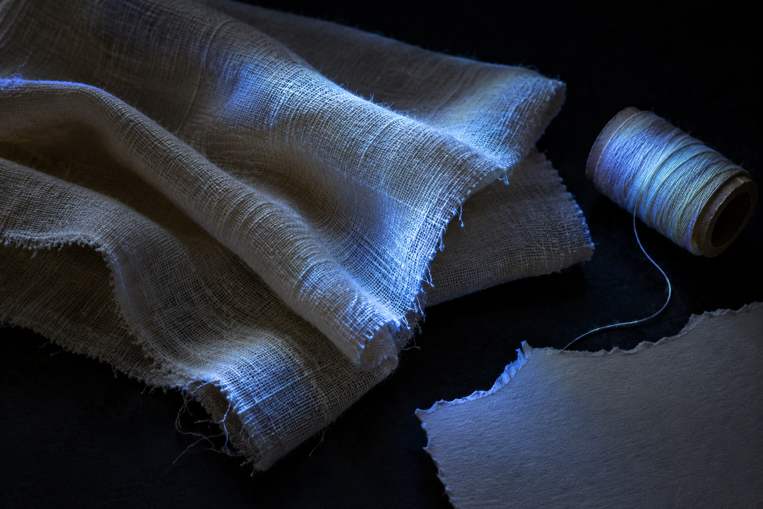
A dark background helps weak emission read, but a black-looking fabric or support can respond unexpectedly under the chosen illumination. Test the empty scene as well as the subject. A bright thread, label or patch of dust can become the most emphatic mark in a carefully arranged still life.
Once the scene is controlled, place the luminous areas deliberately. A diagonal stem can lead toward a flower's center; a low arc of liquid can balance the upright shape of a vessel. Leave dark intervals between competing accents. Darkness should separate and describe, not simply conceal every inconvenient part of the object.
Use a reference image under ordinary visible illumination when comparison matters. Keep the framing and relevant settings documented. The paired views answer different questions, so do not imply that an ordinary-light photograph is an inferior version waiting to be replaced by the more colorful one.
Hard detail and soft emission
Sharp focus can preserve the grain of a petal, a fracture edge or individual fibers. That detail makes the object feel present even when its colors seem unfamiliar. A smaller focused region creates intimacy; a deeper plane of sharpness makes a specimen-like arrangement easier to compare. Choose the depth of field for the question the composition asks.
Softness has a different role. Gentle transitions through a glowing liquid or between responsive and unresponsive areas can communicate volume. Excessive bloom spreads every bright region into its neighbors and makes distinct materials look like the same computer effect. Retain a few crisp edges beside the softer transitions.
Expose and process with the brightest colored regions in mind. A frame can look comfortably dark while one color channel has already lost detail. Inspect the recorded channels rather than relying only on the overall impression. Keep white-balance and processing choices consistent when comparing captures, and disclose major creative changes.
From conservation evidence to botanical images
The Library of Congress documents UVA-induced visible fluorescence as a method for differentiating materials. Smithsonian conservators describe using fluorescence imaging in their study of pichwai paintings, where it can help reveal repairs and suggest material differences. Such observations guide investigation; a color in a photograph is not a conclusive pigment identification.
Photographer Craig P. Burrows offers a contemporary artistic reference through his botanical UVIVF practice. His own explanation makes an equally useful distinction: these pictures do not reproduce exactly what bees see. Recording visible emission from UV excitation is different from recording reflected UV, and neither alone reproduces an animal's complete visual experience.
Fluorescence also differs from bioluminescence, where light is produced through a biological chemical process, and from the persistent afterglow associated with phosphorescence. A glowing-looking AI picture does not establish which process a real subject would exhibit.
Five visual problems to explore
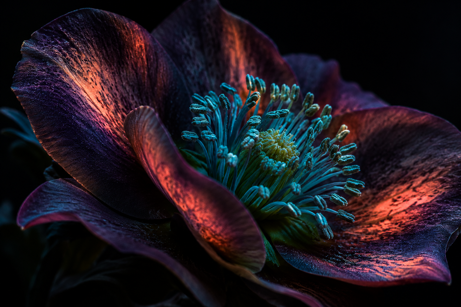
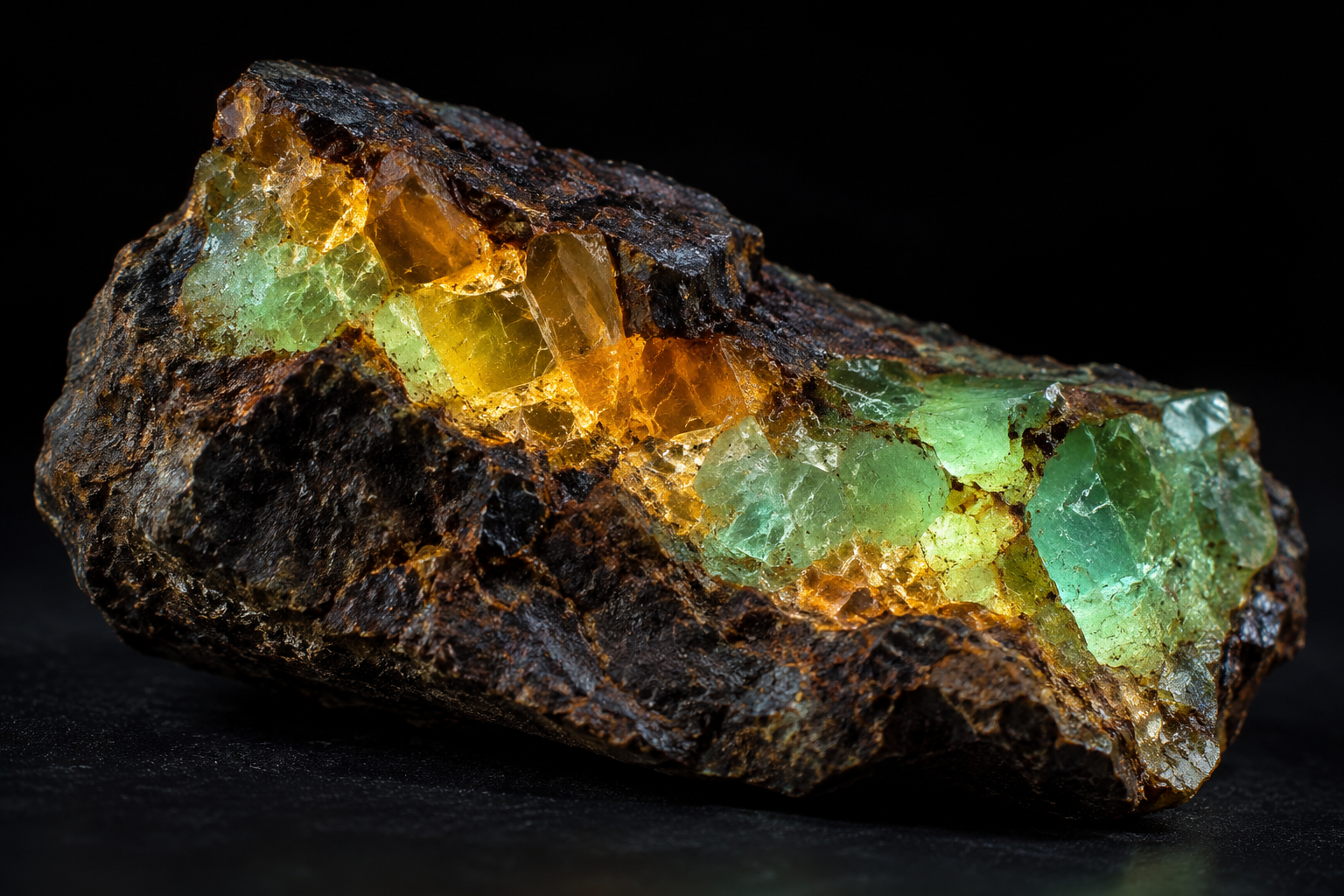
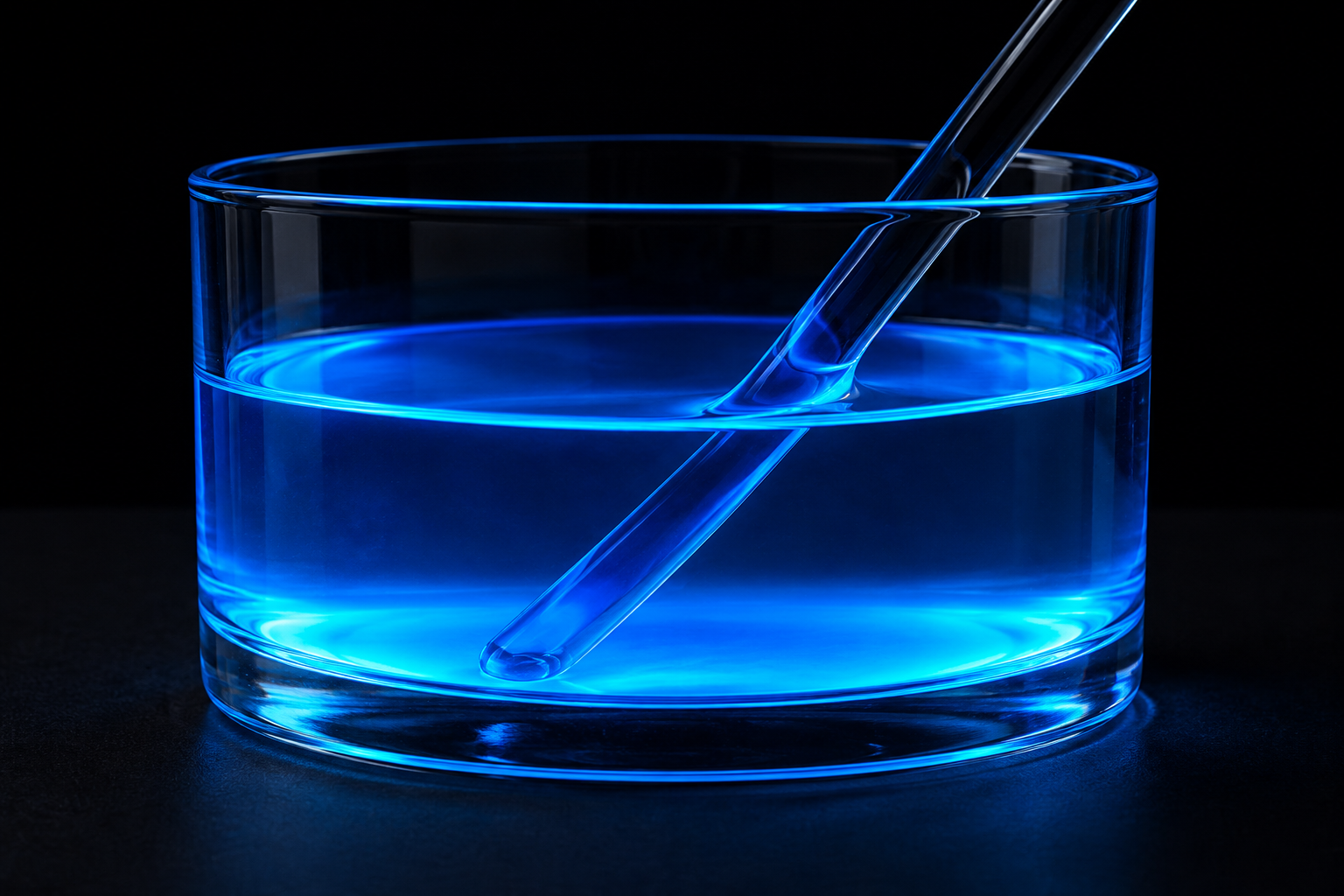
A flower macro tests whether selective brightness can direct the eye through a complex organic form. A mineral study tests the contrast between a luminous interior and a dark, rough exterior. A vessel of fluorescent liquid asks whether color can describe transparent volume without relying on a bright rim. The generated liquid study shows how quickly bright glass boundaries can compete with the colored volume; its graphic treatment is an illustration, not proof that reflected light was excluded.
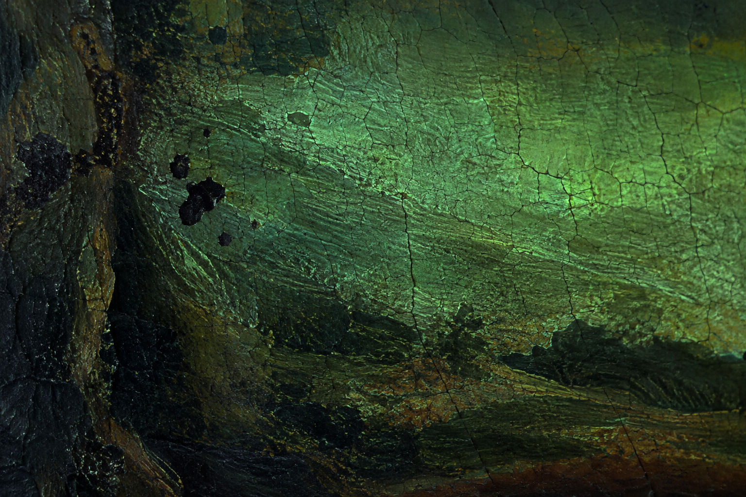
A textile-and-paper still life makes a familiar surface strange while keeping fibers and folds legible. An imagined conservation detail reverses the usual appetite for spectacle: muted greenish emission and dark interruptions can be more informative visually than a rainbow.
The seven AOI images accompanying this entry are original AI-generated interpretations: a dedicated banner, a titled collage and five standalone studies. They illustrate compositional possibilities. Their subjects and palettes are not measured specimens, diagnostic records, or evidence that a particular plant, mineral, pigment or object will fluoresce in the depicted way.
A practical boundary for real UV work
UV is invisible radiation, and exposure can harm eyes and skin. Follow the source manufacturer's controls and use protection specified for the source's wavelength and exposure conditions; do not look into a UV emitter. Keep people and animals out of unnecessary exposure. Germicidal UVC equipment is not a substitute for an appropriate photographic source. The linked CDC guidance explains the risks, while the conservation workflow shows protective goggles as part of its capture procedure.
Generating a visual interpretation needs no physical UV exposure. If the aim is to learn the composition first, use the prompt workshop and compare its imagined results without presenting them as observations.
Positive review — material response becomes composition
AI-authored editorial assessment. At its best, this technique makes material response part of the composition. A dim petal around a bright center, or a luminous seam inside rough stone, gives color a structural purpose. The strongest pictures retain enough texture and darkness to let the viewer recognize the object while discovering a second visual order within it.
Negative review — the glow can overwhelm the evidence
AI-authored editorial assessment. Fluorescence can become a shortcut to spectacle: saturate everything, bury the background and call the result revelation. That weakens both the picture and its claim to observation. Clipped color, excessive bloom, unexplained processing and contamination by visible illumination can erase the distinctions that made the subject interesting. Preserve dark regions, protect surface detail and state how the image was made.
Neutral review — match the claim to the method
AI-authored editorial assessment. UVIVF is a photographic method that can serve scientific documentation, material study or expressive image-making. Those purposes need different controls and captions. A carefully documented capture can support comparison; an openly imaginative illustration can explore its appearance. Neither a striking photograph nor a generated interpretation is, by itself, a chemical identification or a faithful model of another species' vision.
A small poetic detour
Original words from the AOI studio.
Poetry
The light that answers
I brought a lamp the eye could not explain,
and set a small, familiar flower apart.
Its folds replied with islands in the grain,
a quiet map inside the common heart.
The stone kept most of its old darkness whole;
one seam returned a color I could see.
No secret name, no proof of hidden soul—
a different light, a different way to be.
I kept the shadow where the shadow stayed,
and let the bright parts finish what they meant.
The photograph remembers what was made
when borrowed energy was briefly spent.
The prompt workshop
Original creative exercises. These prompts create new illustrations, not documentary evidence.
Let one flower carry the image
Create an original photographic-style interpretation of UV-induced visible fluorescence: one imagined flower against a tested-looking dark background, selective luminous cyan and warm coral details, quieter petals and a restrained stem, crisp surface texture and no visible UV beam. Keep the fluorescence uneven and compositionally purposeful.
This is an AI illustration prompt, not a prediction of a species' fluorescence. Compare selective emission with a uniform neon treatment.
Make the material differences legible
Compose an original UV-fluorescence-inspired still life with a rough dark mineral, one luminous internal seam and a clear vessel containing softly glowing blue liquid. Use negative space to separate the two forms, preserve fracture texture and transparent volume, and avoid bright rim lights that overpower the internal color.
The depicted colors are illustrative. Give each material a different edge behavior instead of using the same glow on every surface.
Reduce the spectacle, retain the question
Imagine a close photographic conservation study of an invented painted panel under UV-induced visible fluorescence. Show muted greenish surface emission, fine cracks and a few dark irregular interruptions, with one sharply focused passage and quiet falloff. No attribution, signature, diagnostic labels or claim of a real artwork.
Use the image to study contrast and surface structure. A generated patch pattern does not establish repairs, age, authenticity or pigment identity.
Your next inspiration
Discover another art style
Follow a visual thread to the next subject.

A smile from the studio
Jokes
The photographer spent an hour arranging one flower for a restrained fluorescence study. The petals were exquisite. The background was perfectly dark. The composition had dignity. Then a stray laundry fiber lit up like the opening ceremony of a small international sporting event. “Please remove the distraction,” said the art director. The photographer pointed to the flower. “Which one? The thread has already asked for a solo exhibition.”
Sources and further exploration
Smithsonian National Museum of Asian Art — how fluorescence helped the study of pichwai paintings