
Scientific imaging method · SEM-inspired artistic interpretation
A scanning electron microscope image reveals surface structure through signals produced by an electron beam. As an artistic reference, its crisp ridges, pores and sculptural tonal contrast can make a tiny form feel monumental. The showcase here contains generated interpretations, not acquired micrographs.
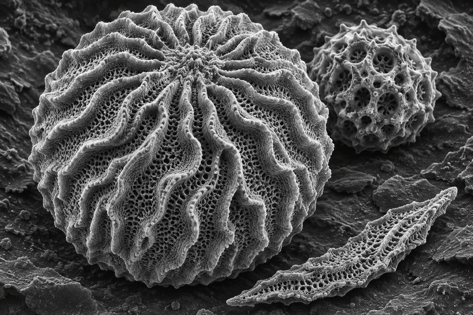
A visual introduction
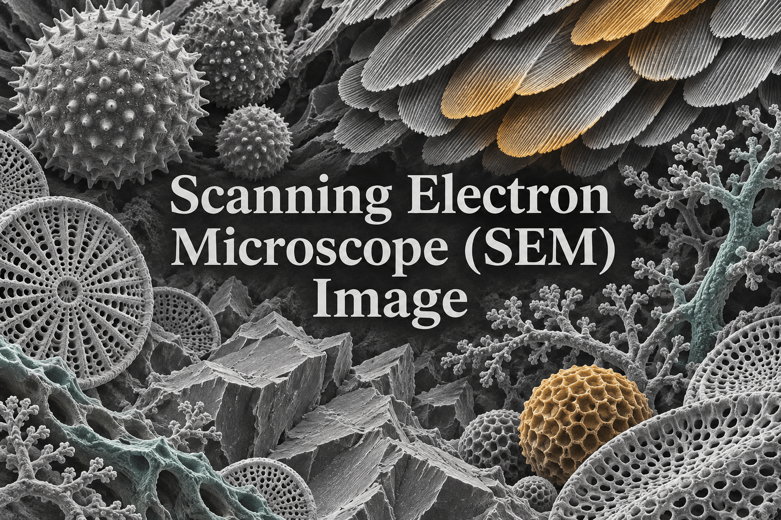
Seven original AI-generated interpretations: a dedicated banner, a centrally titled artistic collage and five standalone studies.
Select an image for a closer look. Use the thumbnails, arrow keys or a swipe to compare examples. Zoom in to inspect details, or share the image that inspires you.
Creative toolbox · take this style further
Choose an input and a destination. Search sends your words to the chosen site; Copy & open puts them on your clipboard to paste there.
Create with AI
Turn your chosen input into an image prompt or a new creative direction.
ChatGPT
Copy your input and paste it into a ChatGPT conversation or image request.
Copy & open ChatGPT (new tab)Claude · Anthropic
Develop an image prompt or explore the style in a new Claude conversation.
Copy & open Claude · Anthropic (new tab)Midjourney
Paste your input into the Imagine bar on the Create page.
Copy & open Midjourney (new tab)Adobe Firefly
Copy your input, open Firefly, and paste it into the prompt field.
Copy & open Adobe Firefly (new tab)Leonardo.Ai
Copy your input, open Leonardo, and paste it into the generation field.
Copy & open Leonardo.Ai (new tab)Image search
Find visual references. Short, concrete keywords usually work best.
Lexica
Search AI images and prompts using your selected input.
Search with this input Lexica (new tab)Pexels
Search the photo and video library. Short keywords often work best.
Search with this input Pexels (new tab)Pixabay
Search photos, illustrations and other images.
Search with this input Pixabay (new tab)Unsplash
Search photographs using the style name or your own keywords.
Search with this input Unsplash (new tab)Wikimedia Commons
Search photographs, artworks and historical images. Check each file for its source and reuse terms.
Search with this input Wikimedia Commons (new tab)Art & research
Explore artists, collections and the ideas behind a style.
Wikipedia
Search for background reading. A style may not have its own article.
Search with this input Wikipedia (new tab)WikiArt
Copy your input into WikiArt’s search to explore artists and artworks.
Copy & open WikiArt (new tab)Europeana
Paste your keywords into the search to explore European museum, library and archive collections.
Copy & open Europeana (new tab)Inspiration
Discover portfolios and visual ideas from creative communities.
Instagram hashtags
Search a hashtag made from your input: spaces and punctuation are removed. Instagram may require sign-in.
Search with this input Instagram hashtags (new tab)ArtStation
Paste your input into artwork search for concept art, environments, characters and visual development.
Copy & open ArtStation (new tab)Behance
Paste your input into project search for illustration, design and visual storytelling.
Copy & open Behance (new tab)A closer look
Organize fine surface detail around a readable silhouette. Use local edge contrast, matte texture, layered ridges and broad areas of focus; distinguish grayscale structure from deliberately assigned false color.
An imaging method, not a single art movement
SEM means scanning electron microscopy or scanning electron microscope, depending on context. It is an imaging method rather than a historical art style. JEOL describes a focused electron beam interacting with the specimen and detectors collecting resulting signals; secondary-electron images are particularly useful for surface topography, while backscattered-electron information can also reveal compositional differences. [1]
This entry treats that imaging language as a visual reference for art. Every gallery image was generated with an image model. None records a specimen placed inside an instrument. A plausible-looking pore, fracture or hair is therefore an invented shape, not evidence of a species, material composition or measured feature.
Why tiny surfaces can feel monumental
A raised edge, repeated pore or fractured plane gives the eye a sequence of spatial clues. When the image removes familiar scale references, a small ridge can suggest a mountain range and a perforated shell can resemble architecture. AOI’s artistic exercise is to preserve that ambiguity while maintaining clear structure.
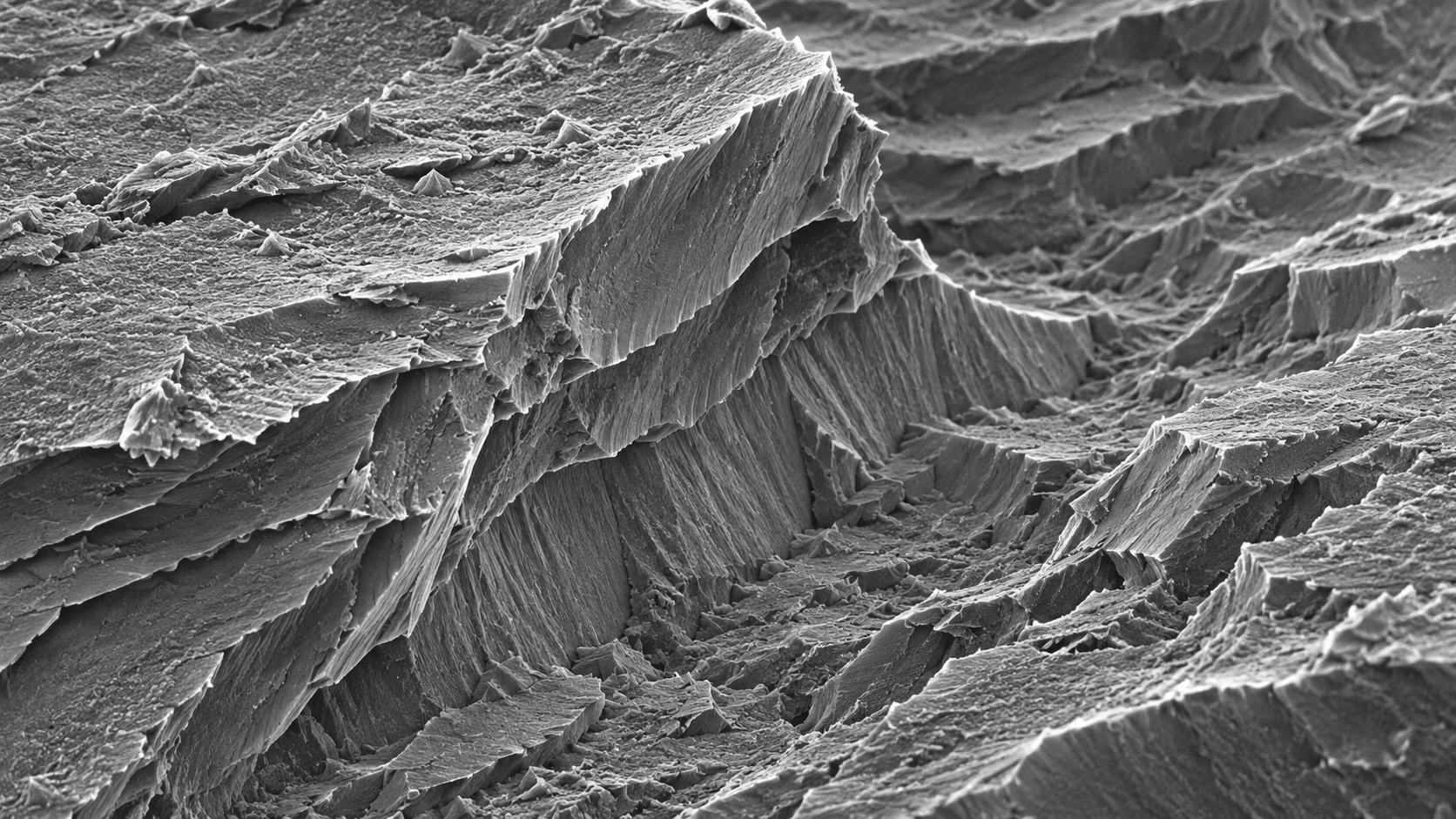
Choose one large organizing form before filling the surface with detail. The dominant grain in the banner provides a silhouette; its ridges supply a middle-scale rhythm; the pores form the finest layer. This hierarchy keeps an intricate image readable as a thumbnail and rewards closer inspection without turning every region into equally busy noise.
Contrast is not ordinary studio lighting
SEM contrast depends on the detected signal and the way the instrument and specimen are arranged. JEOL’s glossary explains that secondary-electron images respond strongly to surface form. [2] A generated illustration can borrow the appearance of bright edges and textured relief, but those marks do not reproduce a detector’s measurements.
For an artistic prompt, ask for local edge emphasis and matte surface variation rather than a metallic shine. Reserve deep darks for recesses and overlapping layers. Large glossy reflections, an optical lens flare or shallow portrait-like bokeh may produce an attractive macro photograph, but they shift the image away from the SEM-inspired vocabulary explored here.
Grayscale and false color
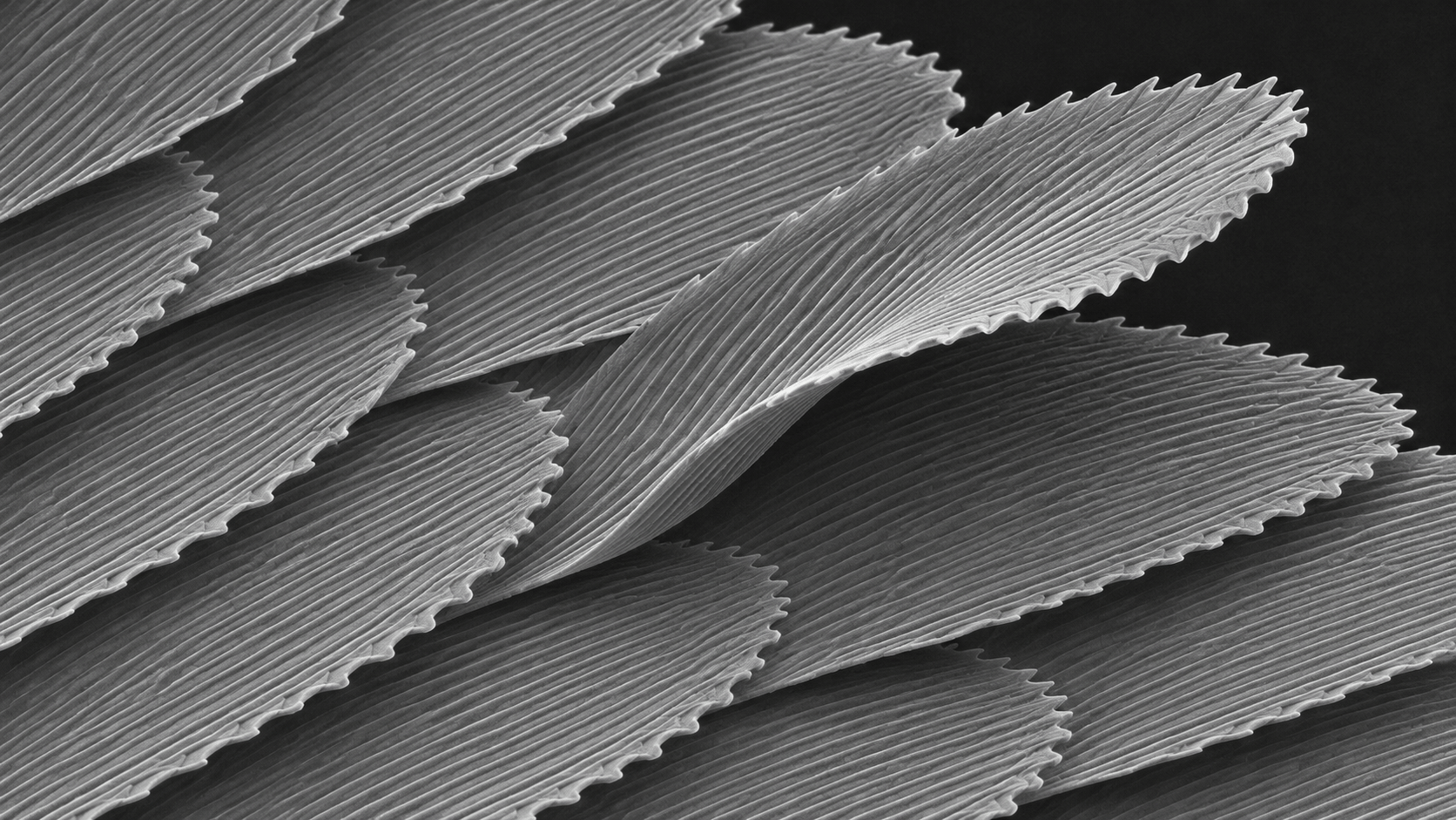
A grayscale study makes tonal structure do the work. The fracture image separates planes through value and texture; the layered-scale study uses repeated ribs and one lifted edge. These are different compositional problems even though both avoid color.
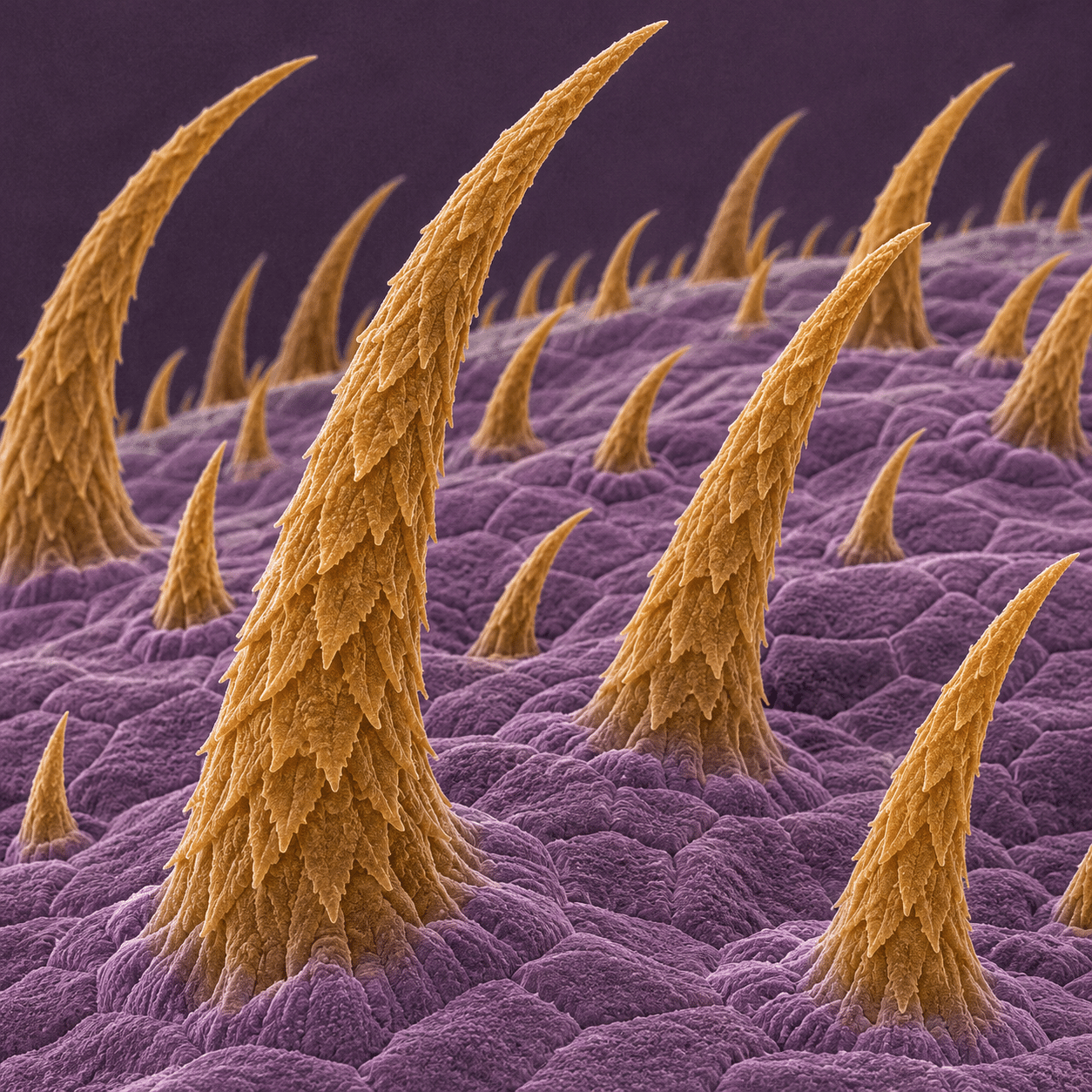
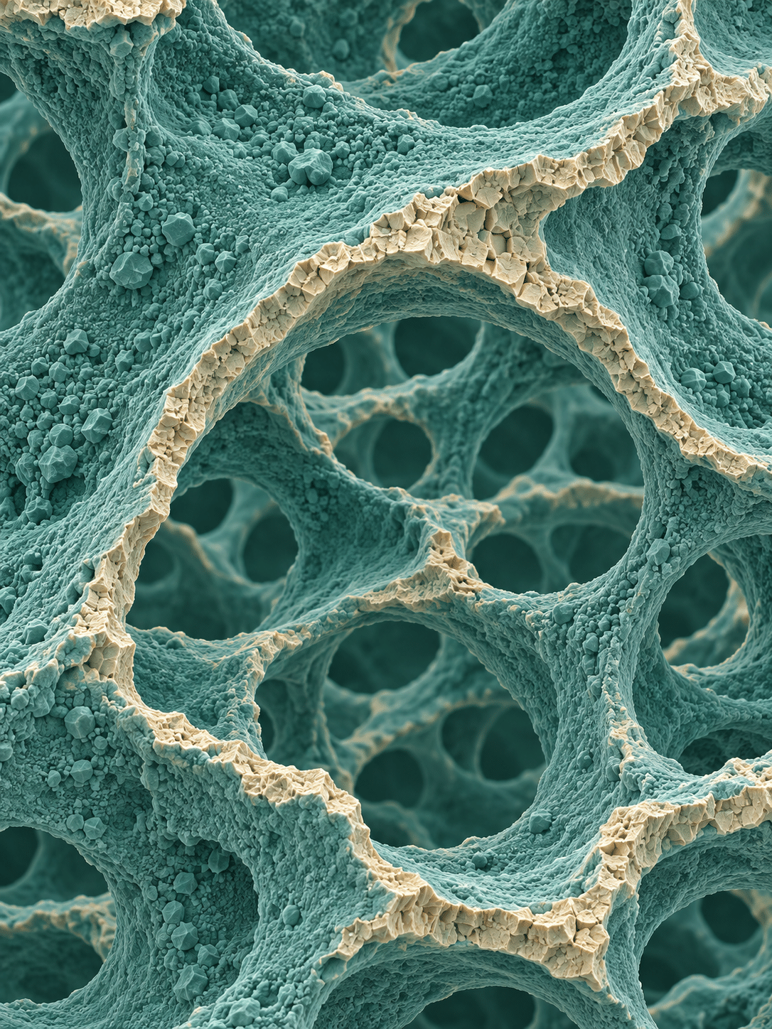
Colorized SEM imagery is also a documented presentation practice. NIST identifies its trichome image as a colorized micrograph and describes another SEM image as having color added for clarity. [3][4] In this generated showcase, amber against purple and teal against ivory are artistic assignments. They do not represent natural specimen colors, elemental maps or calibrated measurements.
Use scientific references without fabricating evidence
When publishing a real micrograph, preserve its source, acquisition context and meaningful scale information. When publishing an invented SEM-inspired artwork, label it accordingly and omit fabricated instrument readouts. A convincing scale bar on a fictional structure would suggest a measurement that never happened.
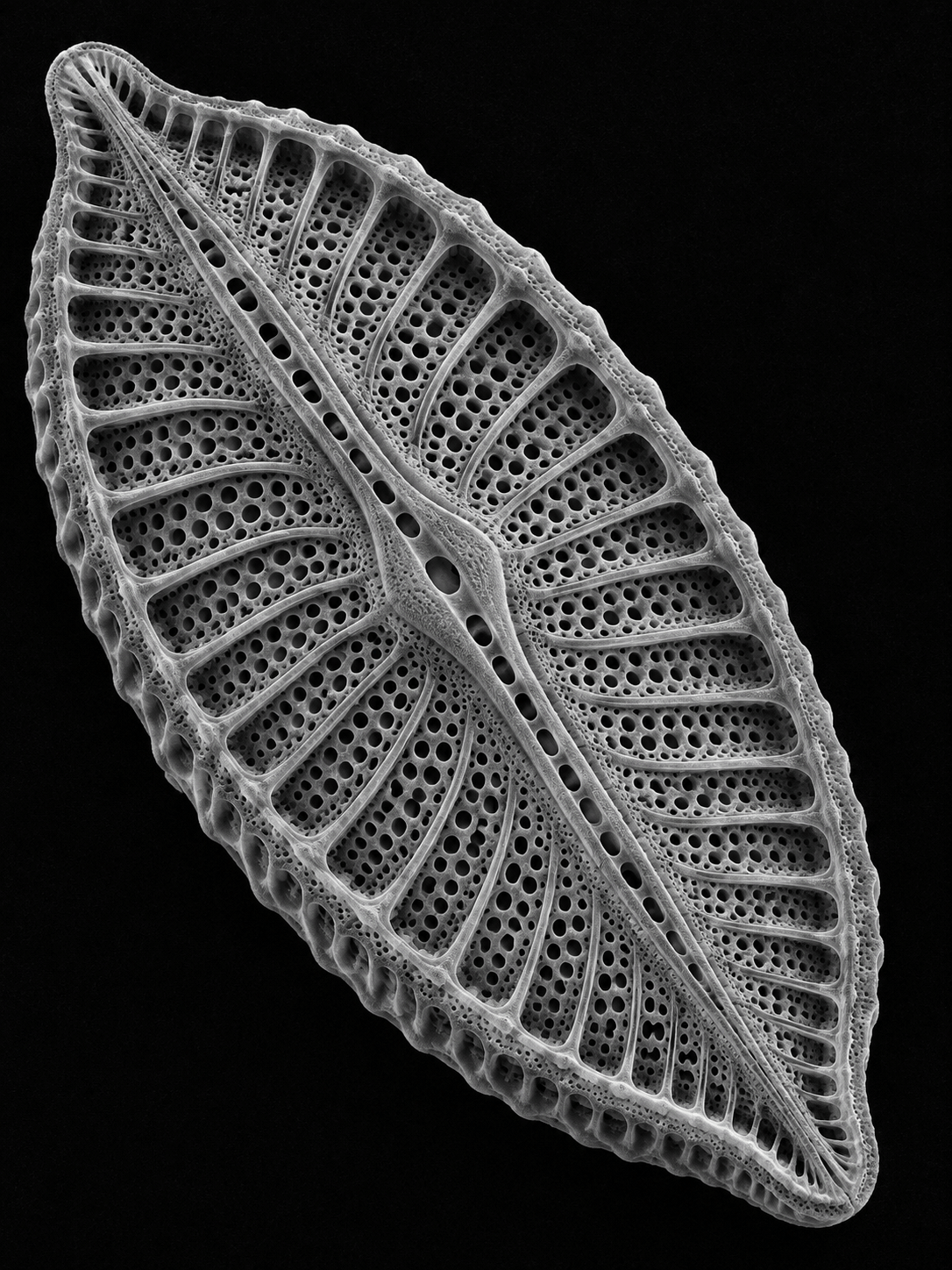
The same care applies to biological names. A generated oval with patterned holes may be diatom-inspired, but resemblance alone cannot establish an organism’s identity. The example captions deliberately describe motifs rather than certifying specimens. The scientific references below explain the method and show real source material; they are not the provenance of the generated gallery.
Five practical directions
Use a porous shell to explore repetition and asymmetry. Use a fracture to organize competing planes. Use a field of hair-like projections to control rhythm and spacing. Use overlapping scales to make one lifted edge interrupt a pattern. Use open-cell architecture to connect foreground and background through holes.
This visual reference suits speculative covers, texture studies and science-inspired illustration when the distinction from evidence remains clear. Avoid the easy formula of adding microscopic noise to any object. Decide what the surface is doing, how the viewer moves through it and which details are essential. The best image needs structure at several scales, not simply the highest possible density.
Positive editorial perspective
AOI editorial perspective (AI-authored). The SEM-inspired approach turns texture into spatial drama. A strong silhouette and nested levels of detail can make pores, ribs and fractures compelling without adding familiar scenic props.
Negative editorial perspective
AOI editorial perspective (AI-authored). Fine noise and artificial edge halos can imitate scientific authority without conveying coherent form. Decorative scale bars make that problem worse. Remove fake measurements and test whether the silhouette and material structure still hold together.
Neutral editorial perspective
AOI editorial perspective (AI-authored). SEM is a scientific imaging method; SEM-inspired art borrows selected visual effects. The generated showcase is useful for composition and texture exploration, but cannot support scientific identification or measurement.
Pocket-sized facts
SEM is an imaging method, not a historical art movement.
Detector signals influence the appearance and interpretation of real SEM images. [1][2]
The gallery uses fictional structures and contains no calibrated scale information.
A small poetic detour
Original words from the AOI studio.
Poetry
A grain becomes a weathered world.
A lifted scale becomes a wing.
The smallest edge unfolds a ridge;
the pores give darkness room to sing.
No traveler stands beside this arch.
No measured mile divides the plain.
The eye invents a distant country
inside the turning of a grain.
Keep wonder close, and labels clear.
Let fact retain its careful name.
A made-up world can still astonish
without pretending how it came.
The prompt workshop
Original creative exercises. These prompts create new illustrations, not documentary evidence.
A sculptural surface
Create an exceptional original SEM-inspired artistic visualization, a fictional generated microstructure rather than an actual scientific micrograph. Portrait-format grayscale fictional diatom-inspired perforated oval shell resting diagonally on black, nested pores and delicate ribs, bold sculptural silhouette, slight natural asymmetry, intricately readable surface. Borrow scanning-electron-image visual grammar: crisp fine surface topology, broad depth of field, directional detector-like edge emphasis, subtle tonal noise, tangible matte microtexture. Avoid glossy CGI, optical lens bokeh, invented measurement markings, instrument readouts and scale bars. No text, labels, numbers, logo, watermark or signature.
Make one silhouette and several scales of texture readable.
A rhythm of layers
Create an exceptional original SEM-inspired artistic visualization, a fictional generated microstructure rather than an actual scientific micrograph. Wide grayscale overlapping wing-scale-inspired plates, parallel fine ribs and serrated edges, rhythmic diagonal fan, one lifted scale gives spatial depth, quiet dark background, not butterfly macro photography. Borrow scanning-electron-image visual grammar: crisp fine surface topology, broad depth of field, directional detector-like edge emphasis, subtle tonal noise, tangible matte microtexture. Avoid glossy CGI, optical lens bokeh, invented measurement markings, instrument readouts and scale bars. No text, labels, numbers, logo, watermark or signature.
Use one raised edge to interrupt the repetition.
Purposeful false color
Create an exceptional original SEM-inspired artistic visualization, a fictional generated microstructure rather than an actual scientific micrograph. Portrait-format false-color porous ceramic-inspired open-cell architecture with an elegant arch of interconnected walls, restrained teal surface and warm ivory fracture edge, intricate granular detail, high depth of field, clearly artificial material study. Borrow scanning-electron-image visual grammar: crisp fine surface topology, broad depth of field, directional detector-like edge emphasis, subtle tonal noise, tangible matte microtexture. Avoid glossy CGI, optical lens bokeh, invented measurement markings, instrument readouts and scale bars. No text, labels, numbers, logo, watermark or signature.
Assign color to distinguish surfaces; do not imply a scientific measurement.
A smile from the studio
Jokes
The grain of dust asked the microscopist for a flattering portrait. “Could you soften the pores?” “That would remove most of the subject.” “Then give me a dramatic mountain background.” The microscopist paused. “You are the mountain background.”
Sources and further exploration
[1] JEOL — how SEM instruments form images and what detector signals can show
[2] JEOL glossary — secondary-electron image and surface contrast
[3] NIST — a documented colorized SEM image of plant trichomes
[4] NIST — carbon nanotube SEM imagery with color added for clarity